Structural and Functional Insights into Emerging SARS-CoV-2 Sub-variant BA.3.2.2/RE.2.2
Under sustained human population immunity, the continuous evolution of Severe Acute Respiratory Syndrome Coronavirus 2 (SARS-CoV-2) generates novel variants characterized by spike (S) protein alterations. Recently, Prof. GAO George Fu’s laboratory at the Institute of Microbiology, Chinese Academy of Sciences (IMCAS), elucidated the structural and functional properties of the surging sub-variant BA.3.2.2 (represented by RE.2.2). The study reveals that RE.2.2 exhibits enhanced human receptor binding, distinct antibody escape patterns, and an unprecedented N-linked glycosylation site at residue N529 of the spike protein. This work was published online in the Proceedings of the National Academy of Sciences of the United States of America (PNAS).
The sublineage BA.3.2.2, derived from Omicron BA.3, has shown an upward trajectory across several regions. Using surface plasmon resonance (SPR) and cryo-electron microscopy (cryo-EM), the researchers determined that the receptor-binding domain (RBD) of RE.2.2 displays high affinity for human angiotensin-converting enzyme 2 (hACE2). Structural analyses revealed that a reverse mutation, R493Q, forms an additional hydrogen bond with hACE2 residue K31, serving as a primary determinant for heightened receptor engagement. Evaluation across diverse animal ACE2 orthologs indicated that RE.2.2 maintains a stable host species spectrum comparable to representative Omicron variants.
Notably, pseudovirus neutralization and binding assays demonstrated a remodeled immune evasion landscape. While RE.2.2 evades multiple antibody classes, several broad neutralizing antibodies that had lost activity against earlier Omicron lineages (such as S2K146 and L4.65) regained potent neutralization against RE.2.2. Cryo-EM ternary structures showed that the key substitution G446D directly facilitates antibody interactions, illustrating that viral antigenic evolution is subject to structural constraints rather than boundless expansion.
Furthermore, comprehensive glycoproteomic profiling via liquid chromatography-tandem mass spectrometry (LC-MS/MS) and cryo-EM revealed up to 26 N-linked glycosylation sites on the S trimer. Most prominently, RE.2.2 harbors a novel N-linked glycosylation at position N529 on the RBD, an alteration previously unobserved in SARS-CoV-2 variants. This modification forms inter-protomer hydrogen bonds that structurally stabilize the spike in a closed conformation, balancing high receptor affinity with regulated viral entry. The findings also validated the "O-follow-N" glycosylation rule, underscoring coordinated glycan organization in coronaviruses.
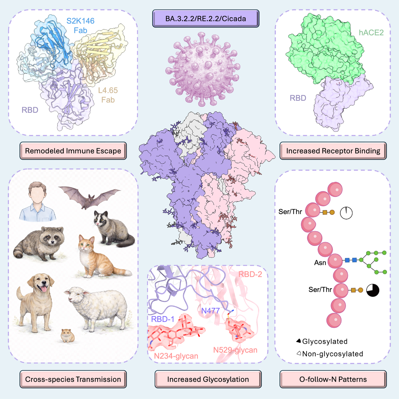
Figure: Structural features, receptor recognition, and glycosylation profiling of SARS-CoV-2 sub-variant BA.3.2.2/RE.2.2. (Image by Prof. GAO George Fu's group)
The study was conducted by Prof. GAO George Fu’s group at IMCAS in collaboration with the University of Chinese Academy of Sciences (UCAS), Shandong First Medical University, Liaoning University, Shandong University, University of Science and Technology of China, and other collaborating institutions. The work was supported by the National Key Research and Development Program of China, the External Cooperation Program of Chinese Academy of Sciences, and the National Natural Science Foundation of China.
Full text link: Increased receptor binding and spike glycosylation, remodeled immune escape of surgingSARS-CoV-2subvariant BA.3.2.2/RE.2.2/Cicada
 Appendix:






